1

Evaluation of vitamin B12 levels and Hypersegmented Neutrophils in pregnant women attending Rivers State University Teaching Hospital, Port Harcourt

Ibiere Allwell Pepple, Catherine Omo Osin and Serekara Gideon Christian*

Department of Medical Laboratory Science, Rivers State University, Nkpolu-Oroworukwo, Port Harcourt, Nigeria All Correspondences to: serekara.christian1@ust.edu.ng

ABSTRACT

This study was aimed at evaluating levels of vitamin B12 and the presence of hypersegmented neutrophils in pregnant women attending Rivers State University Teaching Hospital. It is a comparative and case control study designed to evaluate the levels of vitamin B12 and presence of hypersegmented neutrophils in pregnant women. The study comprises of apparently healthy women in three groups of twenty-five females each (pregnant women who have not had miscarriage, pregnant women with previous miscarriage, and control subjects who have never been pregnant), aged between 30 to 35 years. The study was carried out from July through August 2019. Vitamin B12 levels were determined using ELISA method. Thin films were made and stained using Leishman stain to identify presence of hypersegmented neutrophils. Data was analyzed using Graph Pad prism 8.0.2 statistical package; p<0.05 was considered statistically significant. The results showed significant increase of vitamin B12 in women with miscarriages (193.78±110.63μg/day), when compared to that of women without miscarriages (174.80±53.14 μg/day), and that of non- pregnant women (98.03±9.50 μg/day). Hypersegmented neutrophils were found to be high among women with miscarriages (16%) when compared to those without miscarriage (4%), and control (0%). The miscarriages recorded among the women could be as a result of the presence of hypersegmented neutrophil which is indicative of the tendencies towards miscarriages. However, the high significant level of vitamin B12 in pregnant women with miscarriages may be due to their sea food diet despite the presence of hypersegmented neu trophils; which requ ires fu rtherinvestig a tion.

Keywords: Vitamin B12, Hypersegmented Neutrophils, Pregnancy, Miscarriage
INTRODUCTION

Pregnancy is the period from conception to birth. Pregnancy begins with the fertilization of an ovum (egg) and its implantation. The egg develops into the placenta and the embryo grows to form the foetus. Most eggs implant into the uterus upon fertilization by sperm cell. A normal pregnancy last around 40 weeks from the first day of the woman’s last menstrual period. Normal pregnancy consists of three trimesters of 3 months each (1). Miscarriage (biochemical pregnancy loss) is the pregnancy loss, which occurs after a positive urinary or serum human chorionic gonadotropin (hCG), but before ultrasound or histological detection of pregnancy (<6 weeks) (2). It can also be said to be the loss of the fetus before the 24th week of pregnancy or viability (the ability of the fetus to survive outside the uterus without artificial support). Majority of miscarriages occurs in the first trimester and may be mistaken sometimes for a late menstrual flow (1).

Vitamin B12 (cobalamin or cyanocobalamin) is a water- soluble vitamin that plays a vital role in the activities of several enzymes in the body. It is important in the production of red blood cells in the bone marrow and in the utilization of folic acid and carbohydrate in the diet and the functioning of the nervous system (1). Vitamin B12 sources for humans is food of animal origin. The highest amounts are found in liver and kidney (up to 100μg per 100 g), but it is also present in shellfish, organ and muscle meats, fish, chicken and dairy products (eggs, cheese and milk) in small amounts (6μg/L). Vegetables, fruits and all other foods of non-animal origin are free from cobalamin unless they are contaminated by bacteria. Cooking does not usually destroy cobalamin(3). Vitamin B12 maintains normal folate metabolism which is essential for cell multiplication during pregnancy.

Neutrophil hypersegmentation can be defined as the presence of neutrophils whose nuclei have six or more lobes or the presence of more than 3% of neutrophils with at least five nuclear lobes( 5 ). The presence of hypersegmented neutrophils is suggestive of cobalamin or folate deficiency( 6 ) . So pregnant women with hypersegmented neutrophils correlates with the low amount of vitamin B12 which is detrimental to red blood cell formation and a likely predisposition to neural tube defect and also pregnancy loss (miscarriage). The presence of hypersegmented neutrophils in pregnant women is also an indication of megaloblastic anaemia (5), which puts the pregnancy at risk of a miscarriage. According to Tavasoli et al., the presence of hypersegmented neutrophils indicates low levels of vitamin B12 which results in pregnancy loss due hypercoagulable states and bleeding (7).

In Sub-Saharan Africa, Nigeria inclusive, iron and folate deficiencies were reported to be the most common causes of anaemia in pregnant women (8) Lack and/or insufficient levels of iron supplementation was also reported to be among the most significant risk factors for anaemia to occur during pregnancy(9,10). An anaemic pregnant woman by implication of her condition lacks enough red blood cells for normal metabolic activities and that of the foetus. The resultant effect is that the foetus is deprived of adequate supply of the necessary nutrients required for foetal development.

There is paucity of scientific research information on the levels of vitamin B12 and presence of hypersegmented neutrophils in pregnant women in Rivers State, Nigeria, even though so much effort has been made to improve on the amount of vitamin B12 taken by pregnant women during their routine ante-natal clinic visits. This study was therefore undertaken to ascertain if levels of vitamin B12 and hypersegmented neutrophils could cause pregnancy loss or miscarriage in pregnant women. Hence the need to give this claim a scientific backing.

The aim of the study was to estimate the levels of vitamin B12 and the presence of hypersegmented neutrophils in pregnant women attending River State University Teaching Hospital. The specific objectives of the study are: To estimate the levels of vitamin B12 among pregnant women attending River State University Teaching Hospital; To estimate the number of hypersegmented neutrophils in women attending River State University Teaching Hospital; To ascertain the relationship in values of vitamin B12 estimation with the presence of hypersegmented neutrophils in pregnant women and non- pregnant women.

MATERIALS AND METHODS

Study Design

This is a comparative and case control study which is designed to assess and evaluate the levels of vitamin B12 and the presence of hypersegmented neutrophils in pregnant women attending River State University Teaching Hospital. This study was carried out from July through August 2019.

Study Area

This study was carried out in Rivers State University Teaching Hospital. The hospital is located in Port Harcourt, Rivers State, Nigeria. Port Harcourt is located on GPS coordinates of 4° 49′ 27.0012” N and 7° 2′ 0.9996” E. Rivers State University Teaching Hospital formally Braithwaite Memorial Specialist Hospital (BMSH) is a government-owned hospital, which was named after Eldred Curwen Braithwaite, a British doctor and a pioneer of surgery. It is located in Old GRA, Rivers State. It was established in March 1925.

Study Population

The subjects in this study comprised of apparently healthy pregnant women. Blood samples were drawn from seventy-five (75) women into ethylene diamine tetra acetic acid (EDTA) containers. Their age bracket was 30 to 35 years. They comprised of three (3) groups of 25 subjects each: pregnant women who have not had miscarriage(s), pregnant women that have had previous miscarriage(s) and control subjects who are the non – pregnant women. Women who were less than 30 years or more than 35years were excluded from the study. Convenient sampling method was adopted in recruiting participants.

Informed Consent/Ethical Approval

Informed consent was obtained from the pregnant women before their samples were collected upon clearance from the Department of Medical Laboratory Science, Rivers State University.

Eligibility Criteria

Only apparently healthy pregnant women attending Rivers State University Teaching Hospital and apparently healthy non pregnant women were recruited for this study.

Sample Collection and Storage

A total of 3ml of venous blood was collected by venipuncture with the use of vacutainer needle from each subject and added into individualized vacutainer tube containing 0.5ml of 1.2mg/ml dipotassium ethylene tetra- acetic acid (EDTA). The blood samples in EDTA containers were centrifuged to obtain plasma. The plasma obtained was used to analyse for vitamin B12 using an ELISA reader capable of reading absorbance at 450nm. Thin films were also made immediately before the plasma was separated.

Sample Analysis

The parameters that were analysed were vitamin B12 and presence of hypersegmented neutrophils in thin films.

      1. Determination of Vitamin B12 Using Human Vitamin B12 ELISA Kit, CALBIOTECH, Inc., El Cajon,

U.S.A. Lot No VBE5774; Expiry Date: 2020/05

Principle: It makes use of solid phase ELISA methodology based on the principle of delayed competitive binding. Streptavidin coated wells are incubated with extracted vitamin B12 standards, controls, samples and intrinsic Factor-Biotin conjugate at room temperature for 45 minutes. During the incubation, the biotin-labelled intrinsic factor binds to vitamin B12 in the sample, standard or quality control plasma, after the 45-minutes incubation, vitamin B12 enzyme conjugate is added which competes with the vitamin B12 in the sample, standard, or quality control plasma for the remaining sites on the intrinsic factor for an additional 30 minutes. All unbounded conjugates are then removed and the wells are washed, Next, a solution of tetramethylbenzidine (TMB) reagent is added and incubated at room temperature for 15 minutes, resulting in the development of blue colour. The colour development is stopped with the addition of stop solution, and the absorbance is measured spectrophotometrically at 450 nm. The colour intensity is inversely proportional to the amount of vitamin B12 in the sample. The total procedure run time is 1.5 hours.

Procedure: The EDTA blood samples were centrifuged to obtain plasma. All reagents and specimens were allowed to come to room temperature before use. Desired number of coated strips was placed into the holder. 50µl of extracted Vitamin B12 standards, controls and samples was dispensed into appropriate wells. 50 µl of biotinylated intrinsic factor reagent was dispensed into each well. The microplate was shaken gently for 30 seconds to mix. It was incubated for 45 minutes, at room temperature (250oC). 50 µl of enzyme conjugate was added into all the wells. The microplate was gently shaken for 30 seconds to mix. It was incubated for 30 minutes at room temperature (250oC). The contents were briskly shaken out of the wells. The wells were rinsed 3 times with wash buffer. The wells were stroked sharply on absorbent paper to remove residual water droplets. 100 µl of tetramethylbenzidine (TMB) substrate was dispensed into each well which resulted in the development of a blue coloured solution. The absorbance was read spectrophotometrically at 450nm.

Procedure for Thin Blood Film Preparation

One micro litre of blood was dropped near the end of a slide. The edge of the spreader was placed in front of the blood at an angle of 45o. the spreader was drawn back until it touched the drop of blood and the drop spread along the line of contact between the spreader and the slide on which the film was made. The spreader was moved along the slide with a smooth movement. The film was allowed to air dry. The subject identification was written directly on the frosted end using a lead pencil.

Film Staining using Leishman Staining Technique

Principle: Leishman stain is a mixture of eosin and methylene blue. The acidic dye, eosin variably stains the basic components of the cell which is the cytoplasm and the

basic stain, methylene blue stains the acidic components, especially the nucleus.

Procedure

The slide was placed on the staining rack. The film was flooded with Leishman stain and the stain was allowed to stain for 2 minutes. The stain was later diluted with equal volume of buffered water (pH 6.8) and was allowed to stand for 8 minutes. After 8 minutes, the stain and buffered water was washed off and slide allowed to drain. The back of the slide was then cleaned with cotton wool soaked with 70% alcohol. The film was dried on a rack in a vertical position. The stained f i lmed was examined microscopically using oil immersion objective (100x), ( Olympus microscope) for the presence of hypersegmented neutrophils.

Statistical Analysis

The data generated from this study was analysed and calculated to determine the mean, standard deviation, p- value, f-value using analysis of variance; Tukey’s multiple comparison test was done to check for significance in between groups. Graph pad prism 8.0.2 statistical package was used for the analysis.

RESULTS
    1. Demographic Details of Participants

A total of seventy-five (75) females were recruited for this study. Fifty (50) of the females were pregnant women and were grouped into two (25 were pregnant women that had previous miscarriage(s) and the other 25 were pregnant women that have not had miscarriage(s). Twenty-five (25) women served as control subjects and were non-pregnant. Their age range was between 30-35 years and they were all residents of Port Harcourt, Rivers State. Details are shown in Table 1.

Table 1: Demographic Details of Participants

Age (Years)

No. of Pregnant Women with previous Miscarriage

No. of Pregnant Women without Previous Miscarriage

Control Subjects (Non-pregnant without history of Miscarriage)

30-35 25 25 25

Analysis of Variance of Vitamin B12 in Study Population

Table 2 showed the comparison of vitamin B12 level in pregnant women with previous miscarriage(s), pregnant women without previous miscarriage(s) and non-pregnant women (control)-without history of miscarriage.

Comparatively, the analysis showed that there was statistically significant difference (p<0.05) in the values of vitamin B12 of women with miscarriages (193.78±110.63 μg/day), women without miscarriages (174.80±53.14 μg/day) and the control group (98.03±9.50 μg/day).

Table 2: Comparison of vitamin B12 level using analysis of variance in the study population

Parameter PW+M (A) PW-M (B) Control (C) p-value F-value Inference Tukey’s Multiple Mean±SD Mean±SD Mean±SD Comparison Test

Vitamin 193.78±110.63 174.80±53.14 98.03±9.50 <0.0001 11.95 HS A vs B0.4231 B12 A vs C 0.0001

(µmol/day) B vs C 0.0001

Key: PW+M = Pregnant women with previous miscarriage(s); PW-M = Pregnant women without previous miscarriage(s); HS = Highly significant; SD = Standard deviation. (Applicable to all Tables).

    1. Percentage Distribution of Hypersegmented Neutrophils in Percentage Rate in the Study Population Table 3. shows the percentage distribution of hyper- segmented neutrophils in women with miscarriages, without miscarriages and non-pregnant women. The result showed that the percentage rate of hyper-segmented

neutrophils in women with miscarriages was 4(16%) while the percentage rate of hyper-segmented neutrophils in women without miscarriages was 1(4%). No hyper- segmented neutrophils were found in non-pregnant women.

Table 3: Percentage distribution of hyper-segmented neutrophils in women with miscarriages, without miscarriages and Non-pregnant women

Parameter No. of Women with hyper-segmented Neutrophils Percentage (%)

PW+M (N=25) 4 16
PW-M (N=25) 1 4
Control (N=25) 0 0
4.0 DISCUSSION

From this study, it was observed that there was a significant increase (p<0.05) in the values of vitamin B12 in women with previous miscarriages than those without a history of miscarriage and control participants. Non-pregnant (control) in this study recorded low level of vitamin B12 as a result of them not being on drug supplements and also not having huge appetite for food when compared to pregnant women. The level of vitamin B12 in our study subjects were within the range as reported by VanderJagt et al., (11).

Though it has been reported that maternal vitamin B12 deficiency and increased presence of hypersegmented neutrophils have been associated with increased risk of common pregnancy complications, including spontaneous abortion (miscarriage), low birth weight, intrauterine

among the women could be as a result of the presence of hypersegmented neutrophil which is indicative of the tendencies towards miscarriages. However, the high significant level of vitamin B12 in pregnant women with miscarriages may be due to their sea food diet despite the presence of hypersegmented neutrophils; which requires further investigation.

REFERENCES
  1. British Medical Association. Illustrated Medical Dictionary, 2nd edition, London: Dorling Kindersley Ltd, 2008.
  2. Farquharson RG, Jauniaux E, Exalto N; ESHRE Special Interest Group for Early Pregnancy (SIGEP). Updated and revised nomenclaturefor description of growth restriction and neural tube defects (12, 13). The e a r l y p r e g n a n c y e v e n t s . H u m R e p r o d .

findings of the study indicated that pregnant women with history of miscarriages and those without history of miscarriage had high level of vitamin B12; and so, our findings are at variance with the above reports, probably as a result of the management of the conditions that aforetime may have caused the miscarriages.

The presence of hypersegmented neutrophils is an important diagnostic feature of megaloblastic anaemia; and deficiency of vitamin B12 have been associated with megaloblastic anaemia. In regards to the percentage rate of hypersegmented neutrophils in women with history of miscarriage(s), without history of miscarriage and non- pregnant women, the result showed 4(16%) 1(4%) and 0% in the same order. The hypersegmented neutrophils were found to be high amongst women with history of miscarriage(s) (16%), and this indicates that hypersegmented neutrophils could be associated with miscarriages in some cases in women. The presence of hypersegmented neutrophils in pregnant women with history of miscarriage(s) may probably be only a reflection of their previous miscarriage(s) and tendency towards a miscarriage.

5.0 CONCLUSION

The study revealed that women with miscarriages recorded

2 0 0 5 ; 2 0 ( 1 1 ) : 3 0 0 8 – 1 1 . d o i : 1 0 . 1 0 9 3 /

humrep/dei167.[Pub Med].

  1. Hamid, GA. Clinical Haematology. Retrieved from: https;//www.researchgate.net. Accessed: October 10, 2019.
  2. Van Sande H, Jacquemyn Y, Karepouan N, Ajaji M. Vitamin B12 in pregnancy: maternal and fetal/neonatal effects, a review. Open Journal of Obstetrics and Gynecology. 2013;03(07):599-602. DOI: 10.4236/ojog.2013.37107.
  3. Bain JB. Blood cell morphology in health and disease. Retrieved from https://doi.org/10.1016/B978-0-7020- 6696-2.00005-9. Accessed: February 1, 2021.
  4. Thachil J, Bates I. Approach to the diagnosis and classification of blood cell disorders. Retrieved from: http: https://doi.org/10.1016/B978-0-7020-6696- 2.00023-0. Accessed: February 1, 2021.
  5. Tavasoli B, Tabibian S, Majid G, Souri S, Shams M, Rashidpanah J, Firoozkohi F, Shamsizadeh M, Dorgalaleh A. Extensive Hematoma in a Patient with Hereditary Hypersegmentation of Neutrophils. J

high amount of vitamin B12 and high percentage of

Cell Mol Anesth. 2016;1(3):109-14.

hypersegmented neutrophils. The miscarriages recorded 8. Baker SJ, DeMaeyer EM. “Nutritional anemia: It

understanding and control with special reference to the work of the world health organization,” American Journal of Clinical Nutrition, vol. 32, no. 2, pp. 368–417, 1979.

  1. Aikawa R, Khan NC, Sasaki S, Binns CW. “Risk factors for iron-deficiency anaemia among pregnant women living in rural Vietnam,” Public Health Nutrition, vol. 9, no. 4, pp. 443–448, 2006.
  2. Vivek RG, Halappanavar AB, Vivek PR, Halki SB, Maled VS, Deshpande PS. “Prevalence of Anemia and its epidemiological,” Determinants in Pregnant Women, vol. 5, no. 3, pp. 216–223, 2012.
  3. VanderJagt DJ, Brock HS, Melah GS, El-Nafaty AU, Crossey MJ. Glew, RH. (2007). Nutritional factors associated with anaemia in pregnant women in northern Nigeria, Journal of Health, Population and Nutrition. 2007;25(1):75–81.
  4. Gaber KR, Farag MK, Soliman SET, El-Bassyouni HT, El-Kamah G. Maternal vitamin B12 and the risk of fetal neural tube defects in Egyptian patients. Clinical Laboratory. 2007;53(1-2), 69–75.
  5. Molloy AM Kirke PN Troendle JF. et al., Maternal vitamin B12 status and risk of neural tube defects in a population with high neural tube defect prevalence and no folic Acid fortification. Paediatrics. 2009;123(3),917-923.


CITRODORA OIL: A biosafer alternative to xylene as tissue clearing and dewaxing agent

Akpulu S.P and Hamman W.O

Department of Human Anatomy, Faculty of Basic Med. Scin, Ahmadu Bello University Zaria.

Oladele S.B

Department of Vet.Anatomy, Ahmadu Bello University, Zaria.

Ahmed S.A

Department of Pathology, Faculty of Medicine, Ahmadu Bello University, Zaria, Nigeria All Correspondences to: Akpulu S.P E-mail: petosw2000@yahoo.com

ABSTRACT

Since time immemorial, xylene has been one of the most commonly used tissue clearing and dewaxing agent in histology /histopathology laboratories. However, off late, xylene has been reported to have many toxic effects. Numerous solutions have been suggested as possible alternatives to xylene during tissue processing and staining. Most of these suggested alternatives are at best act like xylene. This study compared the efficacy of Citrodora oil, as a possible alternative to xylene in tissue processing and staining procedures. The study was carried out in the department of Human Anatomy ABU Zaria. The Citrodora oil used was extracted in NARICT Zaria. Two pairs of neutral buffered formalin-fixed brain and liver tissues were histologically processed and stained. One pair was cleared and dewaxed in xylene during the tissue processing and staining, while the other pair was treated similarly in Citrodora oil under the same condition. The paraffin sections were stained with H and E, Gordon and Sweet and Golgi, employing the method of Bancroft and Steven 2008. The section and staining quality was evaluated by direct microscopic observation and graded as described by Kunhua, 2012. Data generated were analyzed using SPSS version 20.0. Results from this study showed a similarity in the efficacy of Citrodora oil and xylene as clearing and dewaxing agent and no significant difference (p≥0.05) in section and staining quality when compared. In conclusion, Citrodora oil can be an effective, eco-friendly, and safer alternative to xylene as a clearing and dewaxing agent in the histology /Histopathological laboratories.

Keywords: Citrodora oil, clearing, staining, xylene and alternative.

INTRODUCTION

Biological tissues have to undergo series of ’tissue clearing agents such as: xylene, toluene, chloroform, acetone, kerosene, diaxane, benzene, petrol, methyl salicylate and cedar wood oil. Most clearing agents are processing’ procedures before they are ready to be examined under the microscope. The various steps of tissue processing include fixing, dehydration, clearing and infiltration. Clearing refers to the process of replacing the dehydrant with a substance that is miscible with the embedding medium. It is one of the most critical steps of tissue processing and largely affects the clarity of the final section and hence the precision of diagnosis.

Clearing agents are used to remove alcohols from the tissue before the tissue can be infiltrated with paraffin wax. Clearing agents are sometimes called “de-alcoholization agents” or ante medium. They act as intermediary between the dehydrating and infiltrating solutions. They are miscible with both solutions and have refractive indices similar to proteins with different levels of toxicity (Kieranan, 2010). Most Histology and Histopathology Laboratories use either aromatic solvents, such as xylene, toluene or aliphatic petroleum distillates for the purpose of clearing and de-waxing in the paraffin histological technique (Ankle and Joshi, 2011). There are many

derivatives of aromatic hydrocarbons such as benzene, while others are derived from natural essential oils such as cedar wood oil and olive oil (Hans et al., 1995).

Xylene has probably been the most commonly used chemical in the histology laboratory despite its hazards. Xylene is an aromatic hydrocarbon consisting of a benzene ring with two methyl substituent (C6H4 (CH 3)2 ). It is expensive, but work well for short time clearing of small tissue blocks. Its high solvency factor allows maximum displacement of alcohol and enhancing paraffin infiltration. (Tardif and Brodeur1992, Carson and Hladik, 2009). Xylene does tend to harden tissues a little, but this does not usually interfere with sectioning qualities.(Kieranan, 2010). Long term immersion of tissue in xylene results in tissue distortions (Visfeldt et al., 1982). Xylene has been reported to affect skin, eyes, nervous system, blood, liver and kidneys of animals exposed to it and it can potentially contaminate the working environment (Ankle and Joshi, 2001).

The need to reduce laboratory hazard has been a challenge.

During tissue processing and staining, most of the clearing agents used are among the most noxious and hazardous chemicals with different levels of toxicity (Dapson and Richard, 2005. Several toxicities believed to be caused by intermediate products of xylene metabolism, such as metylbenzaldehyde have been reported by Indu et al., 2014). These include central nervous system disorders, respiratory depression, abdominal pain, dryness and redness of skin, dermatitis, liver diseases, nephrotoxicity, conjunctivitis, and teratogenic and fetotoxic effects. These are in addition to environmental pollution from unsafe disposal of xylene (Ankle et al., 2011) and tissue distortions as a result of long-term immersion of tissue in xylene (Hans et al., 1995).

There have been several attempts to substitute xylene as clearing agent. Recently, xylene alternatives as clearing agents was developed by mixing vegetable oils such as groundnut oil, palm kernel oil and coconut oil either alone as mixture with other clearing agents (Adeneyi et al., 2016). Orange based oil as clearing agents has also been reported by Rene (2000). Some essential oil such as olive, Clove, Coconut oil and Cedal wood oil has been reported (Hans et al., 1995). However, most of these commercially available xylene alternatives are less effective, more expensive, and are not as readily available as xylene (Gosselin et al., 1984; Amdur et al., 1991; Luna, 1992). To the best of our knowledge, there is little or no report of work on the use of Citrodora oil as xylene substitute in tissue processing. Most of these commercially available xylene substitutes are less effective, more expensive, not readily available and are constitute health hazard as or more than xylene itself (Udonkang et al.,2014).

Citrodora oil is extracted from the Eucalyptus plant which belongs to the; Kingdom: Plantae.Order: Myrtales. Family: Genus: Backhousia. Species: Backhousia citriodora. Citrodora oil is a concentrated hydrophobic liquid containing volatile aroma compounds (Pino et al., 2006). It is extracted from the leaves of Eucalyptus plant and also known as eucalyptus oil. It is an essential oil with a clear, sharp, fresh and very distinctive smell, is pale yellow in color and watery in viscosity (Julia, 995). It has molecular weight of 154.25 and the structural formula of C10H180. The main chemical components of citrodora oil are a- pinene, b-pinene, a-phellandrene, 1,8-cineole, limonene, terpinen-4-ol, aromadendrene, epiglobulol, piperitone and globulol. It has been reported to be nontoxic, nonhazardous, nonflammable, biodegradable and used in aromatherapy (Jean-francois, 2011).

MATERIAL AND METHODS

The essential oil of eucalyptus plants was extracted in National Institute for Chemical Research Technology (NARICT), Zaria by hydro distillation method.

500g of the fresh leaves of eucalyptus and citrus peel was separately weighed and packed into a distillation flask fitted with condensers. Heat was supplied to the flask through a steam generator at constant flow. The essential oil which vaporizes with the steam was condensed into a collecting funnel. The oil was then separated by gravity, dried over anhydrous sodium sulphate, measured, labeled and stored in a brown bottle.

Experimental Protocol

The tissues were taken in pairs. One pair is labelled as Citrodora Tissue and the other as Xylene tissue. The two pairs of brain and liver tissues, 5mmx5mm x3mm thick neutral buffered formalin fixed, were histologically processed simultaneously by dehydration, clearing. Infiltration and embedding. All the tissues were subjected to the same treatment except for the clearing. The Citrodora pair tissue was cleared and dewaxed in Citrodora oil while the Xylene pair tissue was cleared and dewaxed in xylene

Figure I: Eucalyptus plant. Source: Biological Technology Co.Ltd

during the processing and staining respectively. Two paraffin sections of 4 and 8-micron thickness were cut from each of the paired tissue blocks using a rotary microtome (Leica RM2 125 RTS) made in England. Tissues sections sets of 4 microns were stained using Hematoxylin and eosin (H and E) to demonstrate the general tissue structures, Gordon and Sweet for reticular fibers while the brain tissues were stained with Golgi silver stain for nerve fibers. The tissues were dewaxed and cleared with their respective clearing agents during the staining and before cover slipping. The section and staining quality was evaluated by direct microscopic observation and graded as described by Kunhua, 2012. Data generated from the study were expressed as mean plus or minus (±) standard deviation (SD). Student t test was used to compare the efficacy of the Citrodora oil with that of the xylene. Data was analyzed by SPSS 20.0. P value less than or equal to (P ≤ 0.05) were

considered statistically significant.

RESULTS

Table 1.1: Clearing and dewaxing effect of Citrodora oil and xylene on sections and staining quality of liver

Section Quality P value

Citrodora Oil 3.500±1.049

Xylene 4.167±0.753

Staining quality (H and E)

Citrodora Oil 3.333±0.816

Xylene 3.333±0.516

This result indicated there were no statistically significant (P ≥ 0.05) between the Citrodora oil and xylene. At the end of clearing and staining, xylene shows no significant

difference in section quality (4.167±0.753) of liver when compared to Citrodora oil (3.500±1.049).

Table 1.2: Clearing and dewaxing effect of Citrodora oil and xylene on sections and staining quality of Brian

Section Quality P value

Citrodora Oil 3.333±0.816

Xylene 3.500±1.225

Staining quality (H and E)

Citrodora Oil 3.333±1.033

Xylene 3.000±0.894

This result indicated there were no statistically significant (P ≥ 0.05) between the Citrodora oil and xylene. The Brain section and staining quality showed no statistical

significant difference both in section and staining quality.

Photomicrographs of Stained Brain and Liver Tissue Sections

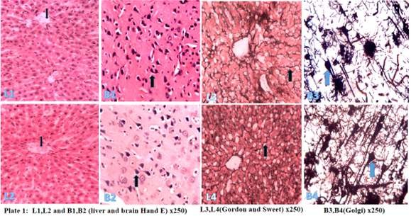
Plate 1: Shows transverse sections of H and E stained liver (L1) and brain (B1) cleared and dewaxed in Citrodora oil and L2 and B2 were cleared and dewaxed in xylene. L3 and L4 are reticular fibers (black arrow) of liver tissue cleared and dewaxed in Citrodora oil and xylene, while B3 and B4 are brain sections demonstrating neurons (blue arrow), were cleared and dewaxed in Citrodora oil and xylene respectively. There was no statistically significant difference across the groups both in the section and staining quality. (H and E X 250).

DISCUSSION

The results of photomicrographs from the present study showed no significant difference in section and staining quality. The present study agrees with the work of Rasmussen et al (1992) on the use of vegetable oils mainly olive and coconut oils instead of xylene in tissue processing, where it was stated that the xylene processed tissues and the vegetable oils’ processed tissues showed only minor or insignificant difference in staining and section quality. According to Kinast (2003), most vegetable oils have higher viscosity between 2 and 5.7 cp at 28% which is reduced by transesterification processes. This may be the reasons for most of the clearing effect on the section and staining qualities observed with some vegetable oil when used as tissue clearing agents. Other physical and phytochemical properties may be responsible for the clearing ability of the Citrodora oil in the present study.

CONCLUSION:

This present study concludes that Citrodora oil can clear and dewax Wistar rat tissues during tissue processing and staining as xylene. Also cytoplasmic and nuclear structures as well as reticular and neural fibers and be demonstrated in Wistar rat tissues cleared in Citrodora oil. Therefore, Citrodora oil can be an effective, eco-friendly and safer alternative to xylene as a clearing and dewaxing agent in the histology /Histopathological laboratory.

Recommendations:

Studies using advanced techniques such as immunohistochemistry, Molecular and enzymatic study, Fluorescence, and electron microscopy techniques of the effects of these essential oils in tissue clearing.

Acknowledgment

The authors sincerely thanked the National Institute for Chemical Research Technology (NARICT), Zaria and their staff for the extraction of the oil, and also the department of human anatomy, Ahmadu Bello University, Zaria for the enabling environment to carry out this work.

REFERENCES
  • Adams, R.P. (2001). Identification of essential oil components by Gas Chromatography and quadruple Mass Spectroscopy. Allured Publishing Corporation Carol Stream IL., SSA, pp 1-698.
  • Awolu, O.O, Obafaye, R.O. and Ayodele, B.S. (2013). Optimization (of Solvent Extraction of Oil from Neem ( Azadirachta indica) and Its Characterizations. Journal of Scientific Research & Reports, 2(1): 304-314.
  • Balch, P.A. (2002). Prescription for Nutritional Healing: the A to Z Guide to Supplements. Penguin. p. 233. 978-1-58333-143-9.
  • Bancroft, J.D. and Stevens (2010). Theory and Practice of Histopathological Techniques 3rd ed P 466. Churchhill Livingstone London, New York.
  • Bancroft, J.D. and Gamble, M. (2008). Theory and practice of Histological Technique.4th Edition. Churchill Living Stone. Britain. p. 84-89.
  • Burke, B.E.. Baillie, J.E. and Olson, R.D. (2004). “Essential oil of Australian lemon myrtle (Backhousia citriodora) in the treatment of molluscum contagiosum in children”, Biomed Pharmacother. 58 (4): 245–247.
  • Caboni, P., Sarais, G., Angioni, A., Garcia, A.J., Lai, F., Dedola, F. and Cabras. (2006). “Residues and Persistence of Neem Formulations on Strawberry after Field Treatment” American Chemical Society, 54 (26), 10026 -10032.
  • Chen, C.Y., He, T., Mao, X.L., Friis, T.E., Qin, R.H., Jian, Y.T., (2010). A novel xylene substitute for histotechnology and histochemistry. Biotech Histochem, 85:231–240.
  • Clark, J.E., Luthy, R.V. (1955). “Separation of Xylenes”. Ind. Eng. Chem. 47(2), 250–253.
  • Dapson, J. and Richard, D. (2005). Hazardous Materials in the Histopathology Laboratory, fourth edition, Anatech, Ltd. 2005
  • Doran, J.C., Brophy, J.J., Lassak, E.V. and House, A.P.N. (2001). “Backhousia citriodora
  • Gilles, M., Zhao, J., An, M. and Agboola, S. (2010). “Chemical Composition and Antimicrobial Properties of Essential Oils of three Australian Eucalyptus Species”. Food Chemistry, 119 (2), 731–737.
  • Gottwald, T.R., Graham, J.H. and Schubert, J.S. (2002). Citrus canker: The pathogen and its impact. Plant Health Progress.
  • Hallur, G., Sivramakrishnan; Bhat, S.V. (2002). “Three New tetranortriterpenoids from Neem Seed Oil” American Chemical Society, 65(8), 1177-1179. 15. Haneke, K.E. (2002). Turpentine [8006-64-2] Review of Toxicological Literature Pub.: National Institute of Environmental Health Sciences.
  • Hans, L., Inger, H., Poul, P. and Eva, B. (1995). Non- hazardous organic solvents in the paraffin embedding technique: A rational approach. Histochemistry, 103:260-269.
  • Hayes, A.J. and Markovic, B. (2002). “Toxicity of Australian essential oil Backhousia citriodora (Lemon myrtle). Part 1. Antimicrobial activity and in vitro cytotoxicity”, Food and Chemical Toxicology 40 (4): 535–543.
  • Jim, S. (2007). Xylene free tissue processing: an evaluation of routine use. Royal Victoria Infirmary, Biopsy Laboratory, Histopathology Department, Newcastle upon Tyne, UK
  • Kieranan, J. A. ( 2010). Histological and Histochemical methods. 4th Edition. Scion publishing limited, Oxford.
  • Kunhua,W.,Chuming,F., Tao,L., Yanmel,Y., Xin,Y., Xiaoming,Z., Xuezhong,G., and
  • Rene, J.B. (2000). Mineral oil: the best xylene substitute. The journal of histotechnology, 3(2)
  • Sakira, F. (2000). The evaluation of an alternative clearing agent to xylene. Clinical Laboratory International.


Association of ABO Blood groups and some diseases–Do Blood groups play a biological role?

Francis Ajeneye*

Pathology Department, Blood Transfusion Maidstone and Tunbridge Well Hospitals NHS Trust, TN2 4QJ, U.K

Oladimeji Olofin

Pathology Department, Haematology Maidstone and Tunbridge Well Hospitals NHS Trust, TN2 4QJ, U.K

Christy Chinyere Fredrick

College of Health sciences, Department of Pathology, University of Abuja, Nigeria All correspondence to: Francis Ajeneye f.ajeneye1@nhs.net

ABSTRACT

The link between ABO/Rh and various diseases have generated much interest over the last decades and have shown some inconsistency. Some diseases are definitely associated with ABO. Do blood groups have biological roles? Many literatures had documented an

association between blood groups and diseases, particularly some neoplasm and hematologic disorders. The loss of blood group A and B antigen expression had been widely documented, which is beyond the scope of this communique. There are also awareness in defining bacterial and parasitic receptors which are closely associated to some known blood groups. Finally, recent studies had observed statistical relationship of blood group with SARS-CoV-2, the association with blood group and SARS-CoV-2 remains insubstantial and evidence must be approached with a sound research design.

Key words: Blood group SARS-CoV-2 Malaria Carcinoma

Over the past few years ABO association with diseases had been widely discussed with emerging research on ABO association and diet, personality and socio-economic statues which is beyond the scope of this short communication.(1,2) Few literatures had proving evidence of increase of group A compared to group with some types of cancer. (3) There is convincing study relating to cancer of the stomach, the association more common in blood group A compared to blood group O. More research across the world agreed with the incidence. ABO association with carcinoma of the stomach seem indisputable(3). Similar association had been also be found in cancer of the colon, and the salivary gland(5). The risk of chorioncarcinoma is critical related to the ABO Blood group of the woman and the partner, women of blood group A with a partner of group O seem to have a highest risk whereas women of blood group A with a partner of blood group A has the lowest risk. There have been some proven relationship of early abortion due to anti-P, Anti-P1 and anti-Pk. The anti-P had been shown to be present in the placenta and anti-PP1PK had been shown to be cytotoxic. (4)

Aird and Bentall (5, 6) showed that group O were 20% more likely to develop peptic ulcer than blood group A. There appears to be a true association between ulceration and absence of secretion. It is not surprising that large quantity of ABH substance are found in the gastrointestinal mucosa,

expression well demonstrated in some human malignancies, the loss of A and B antigen expression was found in 21 of 25 oral carcinoma tumour and correlate with tumour lacking A and B gene expression(7)

There appears to be an association with blood group A, thrombosis, high cholesterol and myocardial infarction. Mourant and colleagues.(8) analysed data from the world literature that patient with thromboembolic diseases include a raised proportion of group A . This applied to women taking oral contraceptive, to pregnancy and puerperal women. There is more evidence of raised thromboembolic episodes in A women than O women. Five year studies found out that A1,B and A1B had a high incidence of myocardial Infarction than O blood group.(9) It was suggested that the etiological pathway of cardiovascular diseases may be differ in patients of different ABO groups owing to difference in their rheology and plasma protein activities.(10,11) These studies consisted of Western Europeans ethnic background, the association did not hold for Asian, African-American or children. It has been shown that cholesterol association may be differ by race.

Mourant(5) made a challenging point earlier that blood

group A individual have a higher level of Factor VIII than group O individual. Group O tend to haemorhage rather than thrombose into the arterial walls thus sustaining more tissue damage. There have also been association with other

failure to secrete ABH substance may lead to peptic ulcer.

coagulation factors such vWF, FV and FIX.(12) Many

There is substantial evidence of decrease activity of glycosyltransferases activities with loss of A and B gene

bacteria such as gram negative organism like Escherichia Coli, have known to have chemical moieties on their

surface that mimics blood group antigens. Springer tested vitro Rosette had been described by to be stronger in

bacteria and found that some bacteria strains showed A, B Group A individuals.(17) The main antigenic ligands

and H (O) specificity.(13) Most blood group scientists responsible for both cytoadherence and antigenic

believe that a similar mechanism must operate in human for the production of naturally occurring antibodies. Small pox virus possess an antigen similar to A antigen, humoral resistance may be more effective in patient with blood group B and O who possess anti-A in their plasma.

The Asian and African distribution of A gene supports the theory of a selective disadvantage among individual infected with smallpox virus. In area like China, India, parts of Russia has a relative increase of the B gene. Resistance to several bacterial and viral infection has been associated with blood groups. Patient with Cholera were likely to be of group O and a one-ninth as likely to be a group AB, several other groups have reported similar associations. (14)

The association of malaria and Duffy blood group system was described by Miller(15), red cell lacking Fya and Fyb were shown to be resistant to the invasion of malaria. It has been shown that black people are resistant to infection by P.Vivax. P.Vivax infection do not occur in individual with Fy(a-b-). The ligand for P. Falciparum is different from the P.Vivax. P.Falciparum invades Fy(a-b-) equally to Fy(a+b+). The parasites exploits adhesion ligands on the endothelial (CD36 and ICAM1), red cell rosette (Blood group A and B), CR1/CD35 and platelets rosettes – platelets glycophorin IV (Cd36) (16) CD36 negative group O appears to be common in malaria endemic areas, this might be a co-incidence or a product of malaria selection, there is also a reduced cyto-adhesion in group O individuals. In-

variation are the members of the P. Falciparum Erythrocyte Membrane Protein-1 (PfEMP1). The encoded PfEMP1var2 carries a two-cysteine-signature associated with rosetting and antibodies to the protein avidly stain the pRBC suggesting that FCR3S1.2 var 2 is the dominant var gene expressed in this parasite.(18)

The parasite ligand-host receptor interactions that mediate cytoadherence is therefore critical to improving our understanding of malaria pathogenesis and developing a vaccine to alleviate disease severity. The second mechanism is to evade specific immune responses through antigenic variation. This involves switching the clonal expression of PfEMP1 antigens, by enabling iRBCs to navigate clear of immunoglobulin (IgG) responses that prevent their cyto-adherence and opsonize them for phagocytosis. PfEMP1 variants thus play a critical role in parasite survival and are prime targets for naturally acquired immunity in African children. Repeating this immunity through PfEMP1 vaccination has been difficult to co-ordinate, especially as thousands of diverse.

There have been other reports associated blood groups with Covid-19, the association of Covid-19 infection and blood groups remains intangible and must be approach with vigilance. The research designs and methodologies identified in recent literatures are not robust and

Cite as: Francis Ajeneye*, Oladimeji Olofin & Christy Chinyere Fredrick (2021). Association of ABO Blood groups and some diseases – Do Blood groups play a biological role?

convincing. A detailed research design that identifies dependent and independent variables and adjust for confounding factors could be a way forward. However, Covid-19 infection could be the results of complex interactions of factors that could vary from genetics, behavioural, metabolic, psychological, social status and environmental risk factors, ABO blood type distribution worldwide varies considerably in different races and should be addressed in studies. In addition to inflammatory response associated with Covid-19 infection,(19) we cannot alterations of the blood group ABO gene in oral overlook mechanism purported by earlier studies that suggested ABO blood group profound influence on the haemostasis, a major determinant of plasma levels of Von

squamous cell carcinoma. Int J Cancer. 2004 Mar 20; 109(2):230-7. doi: 10.1002/ijc.11592. PMID:

14750174.

Willebrand Factor (VWF).(20,21) Literatures also defined

blood group O as a risk factor for increased severe bleeding while blood group non-O is a risk factor for thromboembolic events. The risk of VTE is probably related to the level of VWF and factor VIII in non-group O subjects. A, B, and H blood group antigens are expressed on N-glycans of vWF and influence the half-life of the protein (10 hours for group O and 25 hours for non-O subjects).

CONCLUSION

The relationship between ABO blood groups and overall cancer risk still remains unclear but several meta-analyses conducted over decades suggested there is a strong link. The geographic distribution of ABO antigens worldwide is consistent with a survival advantage in malaria among group O individuals; clinical studies had provided supporting evidence of the effect of ABO group on malaria severity; and that recent discoveries suggest biologic mechanisms linking disease pathogenesis to ABO antigen expression. The mechanisms underlying the associations between ABO blood group and Cardiovascular diseases risk remain unclear, however, several studies of evidence support its potential cardiovascular effects. More research is required to explore and understand the association of Blood groups and SARS-CoV-2.

REFERENCES
  • Golding, J. Hicks, P. Butler NR. Blood group and socio-economic class. Nature 1984; 309; 396
  • Beardmore JA, Karimi-Booshehri F. ABO genes are differentially distributed in socio-economic groups in England. Nature. 1983 Jun 9-15;303(5917):522-4
  • Vogel F. Controversy in human genetics. ABO blood groups and disease. Am J Hum Genet. 1970 Jul;22(4):464-75
  • Lopez M, Cartron J, Cartron JP, Mariotti M, Bony V, Salmon C, Levene C. Cytotoxicity of anti-PP1Pk antibodies and possible relationship with early abortions of p mothers. Clin Immunol Immunopathol. 1983 Aug;28(2):296-303.
  • Aird I, Bentall HH, Mehigan JA, Roberts JAF. The blood groups in relation to peptic ulceration and carcinoma of colon, rectum, breast, and bronchus; an association between the ABO groups and peptic ulceration. Br Med J. 1954 Aug 7;2(4883):315–321.
  • Mourant AE, Kopeć AC, Domaniewska-Sobczak K. Blood-groups and blood-clotting. Lancet. 1971 Jan 30; 1 (7692):223–227.
  • Medalie JH, Goldbourt U. Unrecognized myocardial infarction: five-year incidence, mortality, and risk factors. Ann Intern Med. 1976 May; 84(5):526–531.
  • Dintenfass L, Bauer GE. Dynamic blood coagulation and viscosity and degradation of artificial thrombi in patients with hypertension. Cardiovasc Res. 1970 Jan; 4(1):50-60.
  • Mao, Y., Yang, W., Qi, Q. et al. Blood groups A and AB are associated with increased gastric cancer risk: evidence from a large genetic study and systematic r e v i e w. B M C C a n c e r 1 9 , 1 6 4 ( 2 0 1 9 ) . https://doi.org/10.1186/s12885-019-5355-4
  • Gedde-Dahl TW, Jeremic M, Weisert O. Factor V (proaccelerin) concentration in 1016 blood donors. The effects of age, sex, and ABO blood groups. Scand J Clin Lab Invest. 1975 Jan;35(1):25-30
  • Springer GF. Blood-group and Forssman antigenic determinants shared between microbes and mammalian cells. Prog Allergy. 1971; 15:9-77.
  • Barua D, Paguio AS. ABO blood groups and cholera. Ann Hum Biol. 1977 Sep; 4(5):489-92.
  • Miller LH, McGinniss MH, Holland PV, Sigmon P. The Duffy blood group phenotype in American blacks infected with Plasmodium vivax in Vietnam. Am J Trop Med Hyg. 1978 Nov; 27(6):1069-72.
  • Rug M, Cyrklaff M, Mikkonen A, Lemgruber L, Kuelzer S, Sanchez CP, et al. Export of virulence proteins by malaria-infected erythrocytes involves remodeling of host actin cytoskeleton. Blood. 2014; 124:3459–68.
  • Carlson J, Nash GB, Gabutti V, al-Yaman F, Wahlgren M. Natural protection against severe Plasmodium falciparum malaria due to impaired rosette formation. Blood. 1994 Dec 1; 84(11):3909-14. PMID: 7949147.
  • Kirchgatter K, del Portillo HA: Association of severe no cerebral Plasmodium falciparum malaria in Brazil with expressed PfEMP1 DBL1 alpha sequences lacking cysteine residues. Mol Med. 2002, 8: 16-23.
  • Naitza S, Porcu E, Steri M, et al. A genome-wide association scan on the levels of markers of inammation in Sardinians reveals associations that underpin its complex regulation. PLoS Genet 2012; 8: e1002480.
  • Li J, Wang X, Chen J, Cai Y, Deng A, Yang M. Association between ABO blood groups and risk of SARS-CoV-2 pneumonia. Br J Haematol. 2020 [Epub ahead of print]. DOI: https://doi.org/10.1111/ bjh.16797.
  • Gill JC, Endres-Brooks J, Bauer PJ, et al. The effect of ABO blood group on the diagnosis of von Willebrand disease. Blood 1987; 69:1691-5.
  • Dentali F, Sironi AP, Ageno W, et al. Relationship between ABO blood group and hemorrhage: a systematic literature review and meta-analysis. Semin Thromb Hemost 2013; 39:72-82.
  • Hommel M, David PH, Oligino LD: Surface alterations of erythrocytes in Plasmodium falciparum malaria. Antigenic variation, antigenic diversity, and the role of the spleen. J Exp Med. 1983, 157: 1137- 1148. 10.1084/jem.157.4.1137
  • Gao S, Worm J, Guldberg P, Eiberg H, Krogdahl A, Liu CJ, Reibel J, Dabelsteen E. Genetic and epigenetic